Case Presentation
A 42-year-old male presented to our clinic following a workplace accident where he sustained a complex fracture of his right forearm. The injury occurred when a heavy object fell on his arm during construction work.
Initial Assessment
- Patient: 42-year-old male, right-handed
- Mechanism of Injury: Crush injury from falling construction material
- Symptoms: Severe pain, swelling, deformity of forearm
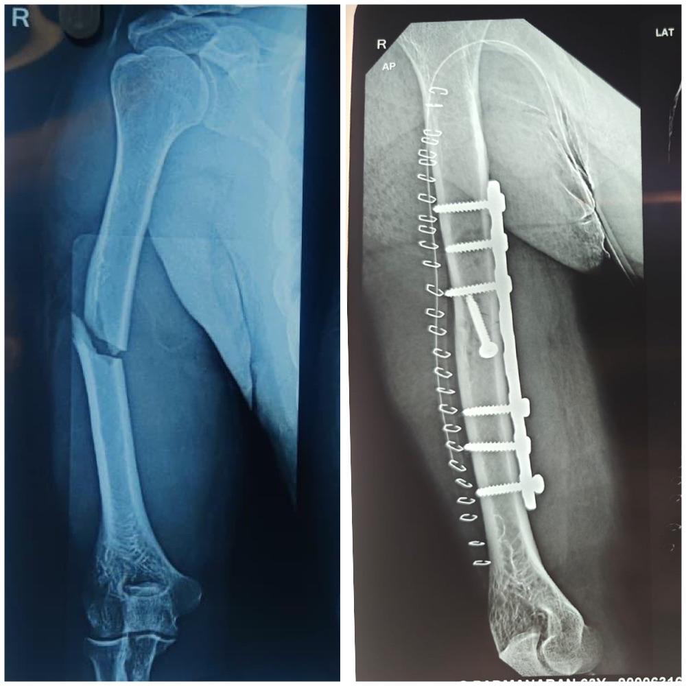
- Initial X-ray: Revealed comminuted fracture of radius and ulna
Diagnosis and Treatment Planning
Radiological Findings
The X-ray examination revealed:
- Comminuted fracture of the distal radius with multiple fragments
- Ulnar shaft fracture with displacement
- Joint involvement with radiocarpal joint disruption
- Soft tissue swelling indicating significant trauma
Surgical Decision
Given the complexity of the fracture and joint involvement, surgical intervention was recommended for:
- Anatomical restoration of bone alignment
- Joint surface reconstruction
- Early mobilization potential
- Prevention of long-term deformity
Surgical Procedure
Pre-operative Preparation
- Medical Clearance: Comprehensive health assessment
- Anesthesia: General anesthesia with regional block
- Positioning: Supine with arm on radiolucent table
- Equipment: Plating system, fluoroscopy, surgical instruments
Surgical Technique
Approach:
- Volar (Henry) approach to the distal radius
- Separate incision for ulnar fixation if needed
- Careful protection of neurovascular structures
Reduction and Fixation:
- Debridement: Removal of hematoma and devitalized tissue
- Fracture Reduction: Anatomical alignment under fluoroscopic guidance
- Temporary Fixation: K-wires for provisional stabilization
- Definitive Fixation: Locking compression plate application
- Bone Grafting: Autograft for bone defects if present
- Closure: Layered closure with drain placement
Intra-operative Challenges
- Fragment Control: Multiple small fragments requiring careful manipulation
- Joint Surface: Precise reconstruction of articular surface
- Soft Tissue: Managing swelling while maintaining exposure
- Stability: Achieving rigid fixation for early mobilization
Post-operative Care
Immediate Post-operative Period
- Hospital Stay: 2 days for monitoring
- Pain Management: Multimodal analgesia protocol
- Antibiotics: 24-hour prophylactic course
- Elevation: Limb elevation to reduce swelling
- Splinting: Temporary splint for soft tissue protection
Rehabilitation Protocol
Week 1-2:
- Wound care and suture removal
- Gentle finger range of motion exercises
- Elbow and shoulder range of motion
- No weight-bearing on injured arm
Week 3-6:
- Initiate wrist range of motion exercises
- Progressive strengthening of forearm muscles
- Light functional activities
- X-ray monitoring of healing
Week 6-12:
- Advanced strengthening exercises
- Grip strength improvement
- Return to light work activities
- Preparation for full activity return
Outcome and Results
Clinical Assessment (3 months)
- Pain: Complete resolution of pain
- Range of Motion: 85% of normal wrist motion restored
- Grip Strength: 90% of contralateral side
- Functional Status: Return to modified work duties
Radiological Assessment
- Bone Healing: Complete union achieved
- Alignment: Anatomical alignment maintained
- Hardware: No signs of loosening or breakage
- Joint Surface: No evidence of post-traumatic arthritis
Patient Satisfaction
The patient reported:
- High satisfaction with surgical outcome
- Return to most daily activities
- Minimal residual symptoms
- Confidence in the treated arm
Key Learning Points
Surgical Considerations
- Anatomical Reduction: Essential for joint function
- Rigid Fixation: Allows early mobilization
- Soft Tissue Management: Crucial for optimal healing
- Rehabilitation: Structured program vital for success
Patient Factors
- Age: Younger patients have better healing potential
- Compliance: adherence to rehab protocol affects outcome
- Occupation: Return to work considerations important
- Expectations: Realistic goals for recovery
Complications and Prevention
Potential Complications
- Non-union: Inadequate bone healing
- Malunion: Healing in poor position
- Infection: Surgical site infection
- Nerve Injury: Temporary or permanent nerve damage
- Complex Regional Pain Syndrome: Chronic pain condition
Prevention Strategies
- Proper Surgical Technique: Meticulous handling of tissues
- Antibiotic Prophylaxis: Infection prevention
- Early Mobilization: Prevents stiffness
- Patient Education: Understanding of rehabilitation importance
Long-term Follow-up
Monitoring Schedule
- 6 weeks: Healing assessment
- 3 months: Functional evaluation
- 6 months: Return to full activity clearance
- 1 year: Final outcome assessment
Expected Long-term Results
- Function: Near-normal wrist function expected
- Strength: Full strength recovery with proper rehab
- Activity: Return to most pre-injury activities
- Hardware: May be removed if symptomatic
This case demonstrates successful management of a complex upper extremity fracture through modern surgical techniques and comprehensive rehabilitation. The outcome highlights the importance of anatomical restoration, rigid fixation, and patient compliance with rehabilitation protocols.
For expert management of complex fractures and orthopedic injuries, contact VPL Ortho and Spine Clinic at +91 9042353157.