Blog Filters

Surgery Case By Dr. Vijaya Anand S on 2026-04-18

Complex Arm Fracture - Successful Surgical Repair

Case Presentation

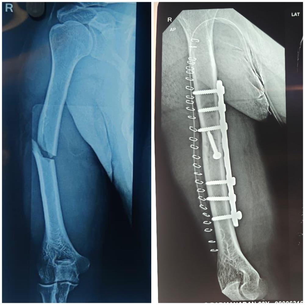
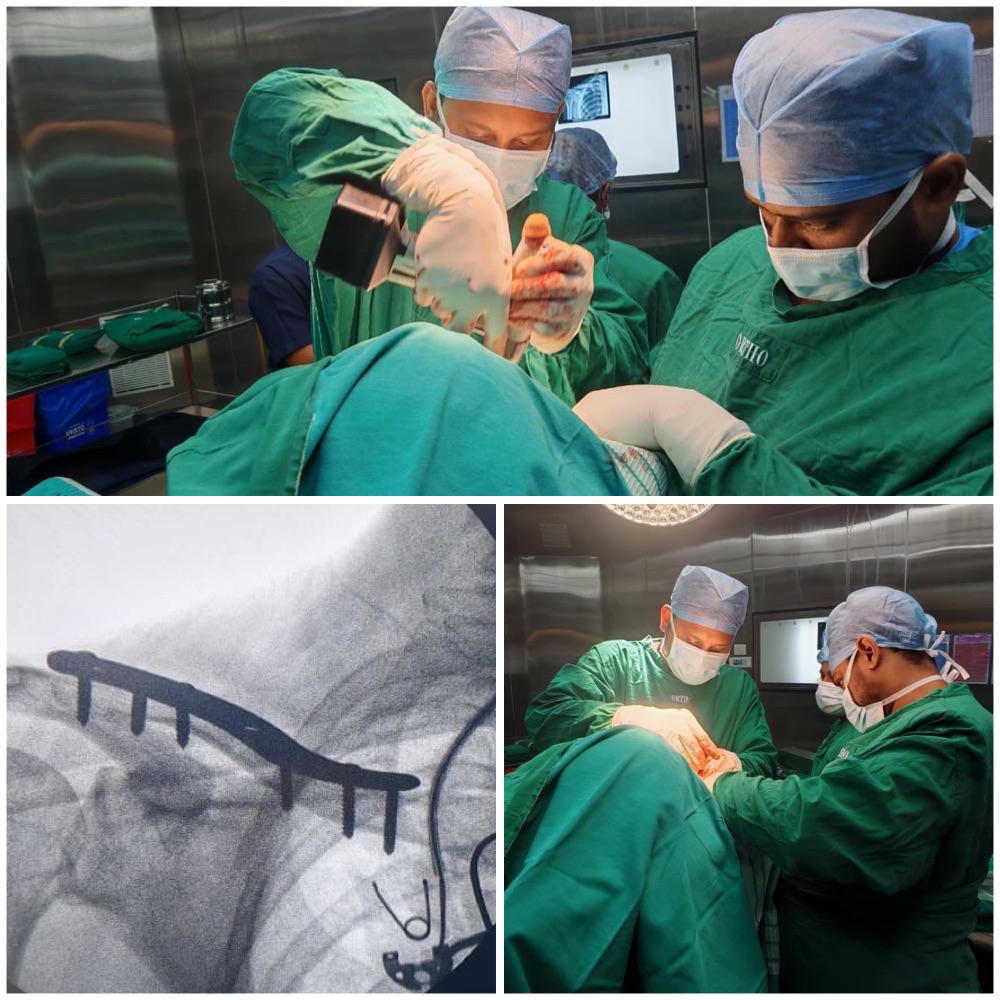
A 42-year-old male presented to our clinic following a workplace accident where he sustained a complex fracture of his right forearm. The injury occurred when a heavy object fell on his arm during construction work.

Initial Assessment

  • Patient: 42-year-old male, right-handed
  • Mechanism of Injury: Crush injury from falling construction material
  • Symptoms: Severe pain, swelling, deformity of forearm
  • Initial X-ray: Revealed comminuted fracture of radius and ulna

Diagnosis and Treatment Planning

Radiological Findings

The X-ray examination revealed:

  • Comminuted fracture of the distal radius with multiple fragments
  • Ulnar shaft fracture with displacement
  • Joint involvement with radiocarpal joint disruption
  • Soft tissue swelling indicating significant trauma

Surgical Decision

Given the complexity of the fracture and joint involvement, surgical intervention was recommended for:

  • Anatomical restoration of bone alignment
  • Joint surface reconstruction
  • Early mobilization potential
  • Prevention of long-term deformity

Surgical Procedure

Pre-operative Preparation

  • Medical Clearance: Comprehensive health assessment
  • Anesthesia: General anesthesia with regional block
  • Positioning: Supine with arm on radiolucent table
  • Equipment: Plating system, fluoroscopy, surgical instruments

Surgical Technique

Approach:

  • Volar (Henry) approach to the distal radius
  • Separate incision for ulnar fixation if needed
  • Careful protection of neurovascular structures

Reduction and Fixation:

  1. Debridement: Removal of hematoma and devitalized tissue
  2. Fracture Reduction: Anatomical alignment under fluoroscopic guidance
  3. Temporary Fixation: K-wires for provisional stabilization
  4. Definitive Fixation: Locking compression plate application
  5. Bone Grafting: Autograft for bone defects if present
  6. Closure: Layered closure with drain placement

Intra-operative Challenges

  • Fragment Control: Multiple small fragments requiring careful manipulation
  • Joint Surface: Precise reconstruction of articular surface
  • Soft Tissue: Managing swelling while maintaining exposure
  • Stability: Achieving rigid fixation for early mobilization

Post-operative Care

Immediate Post-operative Period

  • Hospital Stay: 2 days for monitoring
  • Pain Management: Multimodal analgesia protocol
  • Antibiotics: 24-hour prophylactic course
  • Elevation: Limb elevation to reduce swelling
  • Splinting: Temporary splint for soft tissue protection

Rehabilitation Protocol

Week 1-2:

  • Wound care and suture removal
  • Gentle finger range of motion exercises
  • Elbow and shoulder range of motion
  • No weight-bearing on injured arm

Week 3-6:

  • Initiate wrist range of motion exercises
  • Progressive strengthening of forearm muscles
  • Light functional activities
  • X-ray monitoring of healing

Week 6-12:

  • Advanced strengthening exercises
  • Grip strength improvement
  • Return to light work activities
  • Preparation for full activity return

Outcome and Results

Clinical Assessment (3 months)

  • Pain: Complete resolution of pain
  • Range of Motion: 85% of normal wrist motion restored
  • Grip Strength: 90% of contralateral side
  • Functional Status: Return to modified work duties

Radiological Assessment

  • Bone Healing: Complete union achieved
  • Alignment: Anatomical alignment maintained
  • Hardware: No signs of loosening or breakage
  • Joint Surface: No evidence of post-traumatic arthritis

Patient Satisfaction

The patient reported:

  • High satisfaction with surgical outcome
  • Return to most daily activities
  • Minimal residual symptoms
  • Confidence in the treated arm

Key Learning Points

Surgical Considerations

  1. Anatomical Reduction: Essential for joint function
  2. Rigid Fixation: Allows early mobilization
  3. Soft Tissue Management: Crucial for optimal healing
  4. Rehabilitation: Structured program vital for success

Patient Factors

  1. Age: Younger patients have better healing potential
  2. Compliance: adherence to rehab protocol affects outcome
  3. Occupation: Return to work considerations important
  4. Expectations: Realistic goals for recovery

Complications and Prevention

Potential Complications

  • Non-union: Inadequate bone healing
  • Malunion: Healing in poor position
  • Infection: Surgical site infection
  • Nerve Injury: Temporary or permanent nerve damage
  • Complex Regional Pain Syndrome: Chronic pain condition

Prevention Strategies

  • Proper Surgical Technique: Meticulous handling of tissues
  • Antibiotic Prophylaxis: Infection prevention
  • Early Mobilization: Prevents stiffness
  • Patient Education: Understanding of rehabilitation importance

Long-term Follow-up

Monitoring Schedule

  • 6 weeks: Healing assessment
  • 3 months: Functional evaluation
  • 6 months: Return to full activity clearance
  • 1 year: Final outcome assessment

Expected Long-term Results

  • Function: Near-normal wrist function expected
  • Strength: Full strength recovery with proper rehab
  • Activity: Return to most pre-injury activities
  • Hardware: May be removed if symptomatic

This case demonstrates successful management of a complex upper extremity fracture through modern surgical techniques and comprehensive rehabilitation. The outcome highlights the importance of anatomical restoration, rigid fixation, and patient compliance with rehabilitation protocols.

For expert management of complex fractures and orthopedic injuries, contact VPL Ortho and Spine Clinic at +91 9042353157.

Read More
Complex Arm Fracture - Successful Surgical Repair
Surgery Case By Dr. Vijaya Anand S on 2026-04-18

Complex Spine Surgery - Decompression and Fusion

Case Presentation

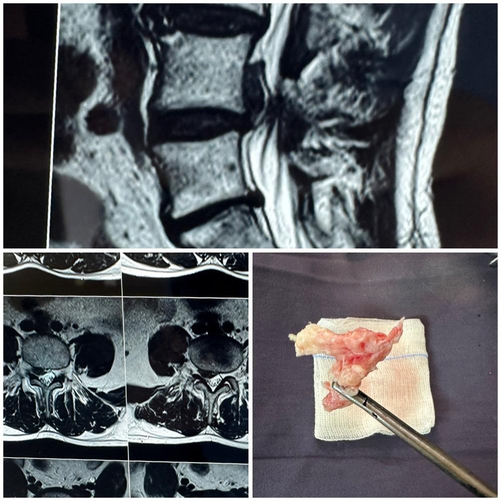
A 62-year-old retired teacher presented with progressive back pain and bilateral leg pain, significantly affecting her ability to walk and perform daily activities. Symptoms had been worsening over 2 years despite conservative treatments.

Initial Assessment

  • Patient: 62-year-old female, retired teacher
  • Chief Complaint: Chronic back pain with bilateral leg pain
  • Duration: 2 years of progressive symptoms
  • Functional Impact: Limited walking distance (<100 meters)
  • Previous Treatment: Physical therapy, medications, epidural injections

Diagnostic Evaluation

Clinical Examination

Physical Findings:

  • Gait: Antalgic gait with limited walking distance
  • Motor Strength: Grade 4/5 in both lower extremities
  • Sensation: Decreased sensation in L4-L5 dermatomes bilaterally
  • Reflexes: Diminished patellar and Achilles reflexes
  • Special Tests: Positive slump test bilaterally

Imaging Studies

X-ray Lumbar Spine:

  • Degenerative Changes: Grade 2 spondylolisthesis at L4-L5
  • Disc Height: Reduced disc height at multiple levels
  • Facet Arthropathy: Severe facet joint degeneration
  • Instability: Dynamic instability on flexion-extension views

MRI Lumbar Spine:

  • Spinal Stenosis: Severe central canal stenosis at L4-L5
  • Foraminal Stenosis: Bilateral foraminal narrowing
  • Disc Herniation: Multiple disc bulges
  • Nerve Compression: Significant nerve root compression

Treatment Planning

Indications for Surgery

Surgical intervention was recommended due to:

  • Progressive Neurological Deficit: Motor weakness
  • Severe Pain: Uncontrolled with conservative measures
  • Functional Limitation: Inability to walk reasonable distances
  • Imaging Correlation: Significant compression matching symptoms

Surgical Plan

Procedure Recommended:

  • L4-L5 Decompression: Laminectomy and foraminotomy
  • Spinal Fusion: Posterior lumbar interbody fusion (PLIF)
  • Instrumentation: Pedicle screw fixation
  • Bone Graft: Autograft with allograft augmentation

Surgical Procedure

Pre-operative Preparation

  • Medical Optimization: Cardiac clearance, anemia correction
  • Anesthesia: General anesthesia with invasive monitoring
  • Positioning: Prone position on spinal table with padding
  • Equipment: Spinal instrumentation set, microscope, neuromonitoring

Surgical Technique

Approach and Exposure:

  1. Incision: Midline incision from L3 to S1
  2. Dissection: Subperiosteal dissection of paraspinal muscles
  3. Instrumentation: Pedicle screw placement at L4 and L5
  4. Verification: Fluoroscopic confirmation of screw placement

Decompression:

  1. Laminectomy: Complete removal of L4 lamina
  2. Foraminotomy: Bilateral foraminotomy at L4-L5
  3. Facetectomy: Partial facetectomy for adequate decompression
  4. Ligamentum Flavum: Removal of thickened ligamentum flavum
  5. Disc Removal: Discectomy at L4-L5 level

Fusion and Reconstruction:

  1. Disc Space Preparation: Endplate preparation and distraction
  2. Cage Placement: Interbody cage with bone graft
  3. Compression: Segmental compression for stability
  4. Rod Placement: Contoured rods connecting pedicle screws
  5. Final Compression: Locking screws in compression

Intra-operative Monitoring

  • EMG Monitoring: Continuous nerve monitoring
  • SSEP: Somatosensory evoked potentials
  • MEP: Motor evoked potentials
  • Wake-up Test: No neurological changes detected

Post-operative Care

Immediate Post-operative Period

  • ICU Care: 24-hour monitoring in ICU
  • Pain Management: Epidural analgesia and PCA pump
  • Mobilization: Ambulation on postoperative day 2
  • Hospital Stay: 5 days for monitoring and rehabilitation

Rehabilitation Protocol

Week 1-2:

  • Bed rest with bathroom privileges
  • Gentle range of motion exercises
  • Deep breathing and incentive spirometry
  • Wound care and monitoring

Week 3-6:

  • Progressive ambulation with walker
  • Core strengthening exercises
  • Back extension exercises
  • Transition to cane for support

Week 6-12:

  • Advanced strengthening program
  • Gait training and balance exercises
  • Activities of daily living training
  • Preparation for return to light activities

Month 3-6:

  • Return to normal activities
  • Driving clearance
  • Recreational activities as tolerated
  • Final functional assessment

Outcome and Results

6-Month Follow-up

Clinical Assessment:

  • Pain: Significant reduction in back and leg pain
  • Function: Walking distance >1 kilometer
  • Motor: Full strength restored (5/5)
  • Sensation: Normal sensation in lower extremities

Radiological Assessment:

  • Fusion: Solid fusion achieved at L4-L5
  • Alignment: Restoration of normal alignment
  • Hardware: No signs of loosening or failure
  • Decompression: Adequate neural decompression confirmed

Patient-Reported Outcomes

  • Pain Score: Reduced from 8/10 to 2/10
  • Functional Status: Return to daily activities
  • Quality of Life: Significant improvement
  • Satisfaction: Very satisfied with surgical outcome

Surgical Considerations

Complex Spine Surgery Challenges

  1. Patient Age: Increased surgical risks in elderly
  2. Medical Comorbidities: Management of associated conditions
  3. Bone Quality: Osteoporosis affecting fixation
  4. Deformity Correction: Restoration of normal alignment
  5. Neurological Risk: Protection of neural structures

Technical Pearls

  1. Meticulous Decompression: Adequate neural element release
  2. Solid Fixation: Proper pedicle screw placement
  3. Fusion Technique: Proper preparation and grafting
  4. Blood Loss Management: Minimizing intraoperative bleeding
  5. Complication Prevention: Proactive complication avoidance

Complications and Management

Potential Complications

  • Dural Tear: Cerebrospinal fluid leak management
  • Nerve Injury: Intraoperative nerve monitoring protection
  • Hardware Failure: Proper technique and patient selection
  • Non-union: Smoking cessation and bone health optimization
  • Infection: Prophylactic antibiotics and sterile technique

Prevention Strategies

  • Pre-operative Optimization: Medical risk factor management
  • Intraoperative Monitoring: Continuous neurological monitoring
  • Proper Technique: Meticulous surgical execution
  • Postoperative Care: Appropriate rehabilitation and monitoring

Long-term Follow-up

Monitoring Schedule

  • 6 weeks: Wound check and initial assessment
  • 3 months: Fusion assessment and functional evaluation
  • 6 months: Final clinical and radiological assessment
  • 1 year: Long-term outcome evaluation

Expected Long-term Results

  • Pain Relief: 70-80% success rate for pain reduction
  • Function: Improved walking ability and daily function
  • Durability: Long-lasting fusion and symptom relief
  • Quality of Life: Significant improvement in overall well-being

Patient Selection Criteria

Ideal Candidates

  • Significant Symptoms: Pain and functional limitation
  • Failed Conservative: Adequate trial of non-surgical treatment
  • Corresponding Imaging: Radiological findings match symptoms
  • Reasonable Health: Appropriate medical fitness for surgery
  • Realistic Expectations: Understanding of recovery process

Contraindications

  • Severe Medical Illness: Uncontrolled comorbidities
  • Active Infection: Systemic or local infection
  • Severe Osteoporosis: Poor bone quality for fixation
  • Non-compliance: Inability to follow postoperative instructions

Rehabilitation Philosophy

Principles of Recovery

  1. Early Mobilization: Prevent complications of bed rest
  2. Progressive Loading: Gradual increase in activity
  3. Core Strengthening: Support for spinal fusion
  4. Patient Education: Understanding of limitations and goals
  5. Long-term Maintenance: Lifetime spine health practices

This case demonstrates successful management of complex spinal stenosis with spondylolisthesis through decompression and fusion surgery. The outcome highlights the importance of proper patient selection, meticulous surgical technique, and comprehensive rehabilitation for optimal results.

For expert evaluation and treatment of complex spine conditions, contact VPL Ortho and Spine Clinic at +91 9042353157.

Read More
Complex Spine Surgery - Decompression and Fusion
Awareness By Dr. Vijaya Anand S on 2026-04-18

Fall Prevention - Protecting Yourself and Your Loved Ones

Why Fall Prevention Matters

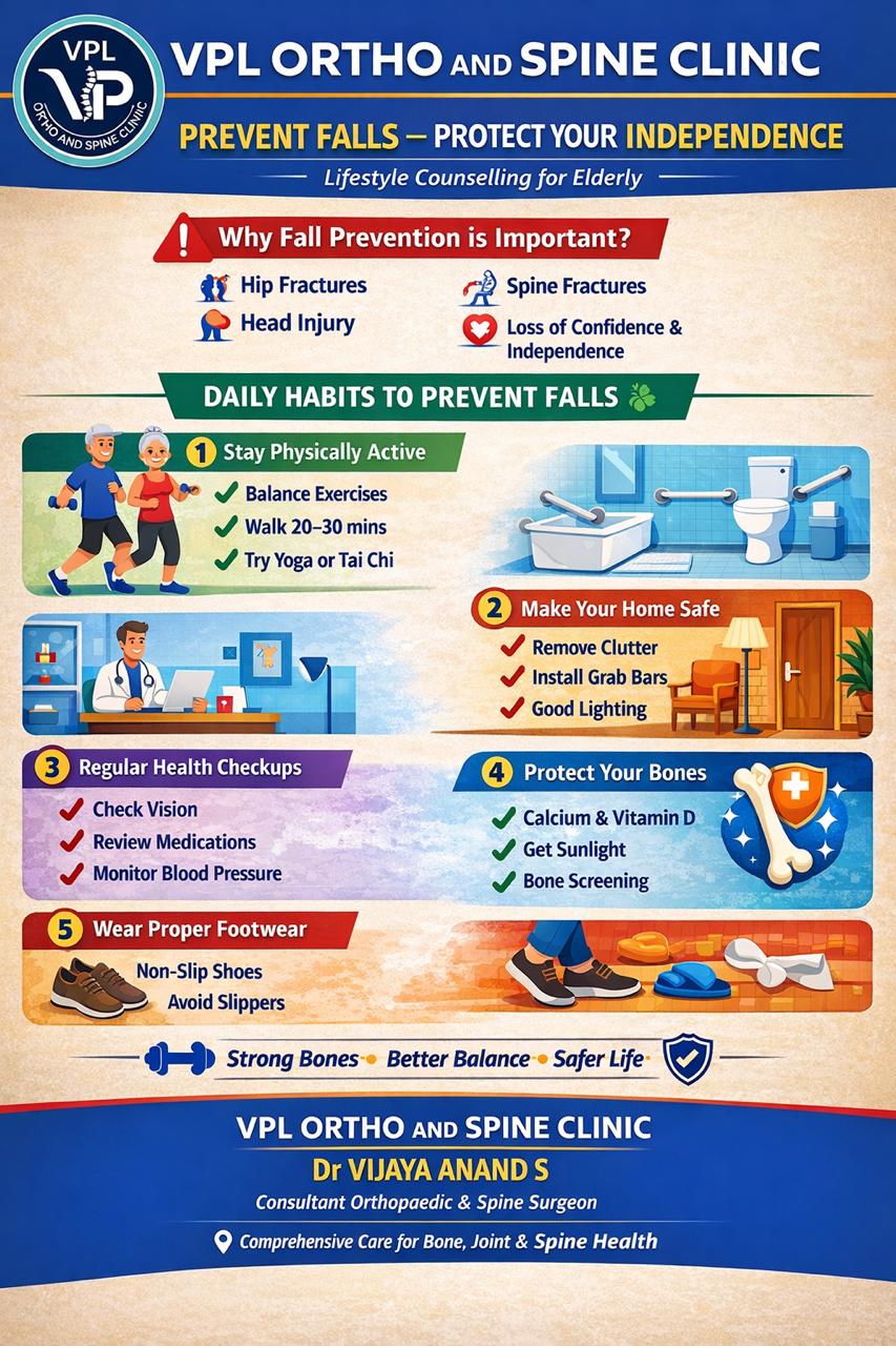
Falls are a leading cause of injury, especially among older adults. According to studies, one in four adults aged 65 and older falls each year. Falls can lead to serious injuries, including fractures, head injuries, and reduced quality of life. The good news is that most falls are preventable.

Who is at Risk?

High-Risk Groups:

  • Adults over 65 years old
  • People with balance or walking problems
  • Those taking multiple medications
  • Individuals with vision problems
  • People with chronic conditions (diabetes, heart disease)
  • Those with previous fall history

Home Safety Modifications

Remove Hazards

  • Clear pathways: Remove clutter, cords, and rugs
  • Secure carpets: Use non-slip backing or double-sided tape
  • Fix uneven surfaces: Repair cracked walkways and stairs
  • Adequate lighting: Install night lights and bright bulbs
  • Bathroom safety: Install grab bars and non-slip mats

Essential Safety Equipment

  • Grab bars: In bathrooms near toilet and shower
  • Handrails: On both sides of staircases
  • Non-slip mats: In bathroom and shower
  • Raised toilet seat: If mobility is limited
  • Shower chair: For those with balance issues

Physical Conditioning

Balance Exercises

  • Tai Chi: Improves balance and reduces fall risk
  • Yoga: Enhances flexibility and stability
  • Standing on one leg: Simple balance exercise
  • Heel-to-toe walking: Improves coordination

Strength Training

  • Leg exercises: Squats, lunges, calf raises
  • Core strengthening: Planks, bridges
  • Resistance training: Using bands or light weights

Flexibility

  • Regular stretching: Maintains range of motion
  • Gentle yoga: Improves flexibility safely

Medical Considerations

Medication Review

  • Consult your doctor: Review all medications regularly
  • Be aware of side effects: Dizziness, drowsiness
  • Time medications: Take when you can rest afterward
  • Avoid alcohol: Can increase fall risk with medications

Vision Care

  • Regular eye exams: At least annually
  • Update prescriptions: Correct vision problems
  • Use proper lighting: Ensure adequate illumination
  • Clean glasses: Keep lenses clean and scratch-free

Foot Health

  • Proper footwear: Supportive, non-slip shoes
  • Avoid walking barefoot: Especially on smooth surfaces
  • Check foot problems: Corns, calluses, nail issues
  • Custom orthotics: If recommended by doctor

Lifestyle Modifications

Daily Habits

  • Get up slowly: From bed or chairs to avoid dizziness
  • Use assistive devices: Canes or walkers if needed
  • Stay hydrated: Dehydration can cause dizziness
  • Regular exercise: Maintain strength and balance

Nutrition

  • Adequate calcium: For bone health
  • Vitamin D: Essential for bone strength
  • Protein: Maintains muscle mass
  • Stay hydrated: Prevents dizziness

What to Do If You Fall

If you fall and can get up:

  1. Take deep breaths to stay calm
  2. Check for injuries before moving
  3. Roll onto your side and push up to hands and knees
  4. Crawl to a sturdy chair
  5. Place hands on chair and slide one foot forward
  6. Slowly stand up

If you fall and can't get up:

  1. Call for help immediately
  2. Try to move to a more comfortable position
  3. Keep warm with nearby blankets or clothing
  4. Don't try to stand if injured

When to Seek Medical Attention

After a fall, see a doctor if you:

  • Hit your head or feel confused
  • Have severe pain
  • Can't move or bear weight
  • Have obvious injuries or bleeding
  • Feel dizzy or nauseous

Remember, fall prevention is a team effort involving you, your family, and healthcare providers. Taking these preventive steps can significantly reduce your risk of falls and maintain your independence.

For personalized fall risk assessment and orthopedic care, contact VPL Ortho and Spine Clinic at +91 9042353157.

Read More
Fall Prevention - Protecting Yourself and Your Loved Ones
Surgery Case By Dr. Vijaya Anand S on 2026-04-18

Microdiscectomy - Relief from Sciatic Pain

Case Presentation

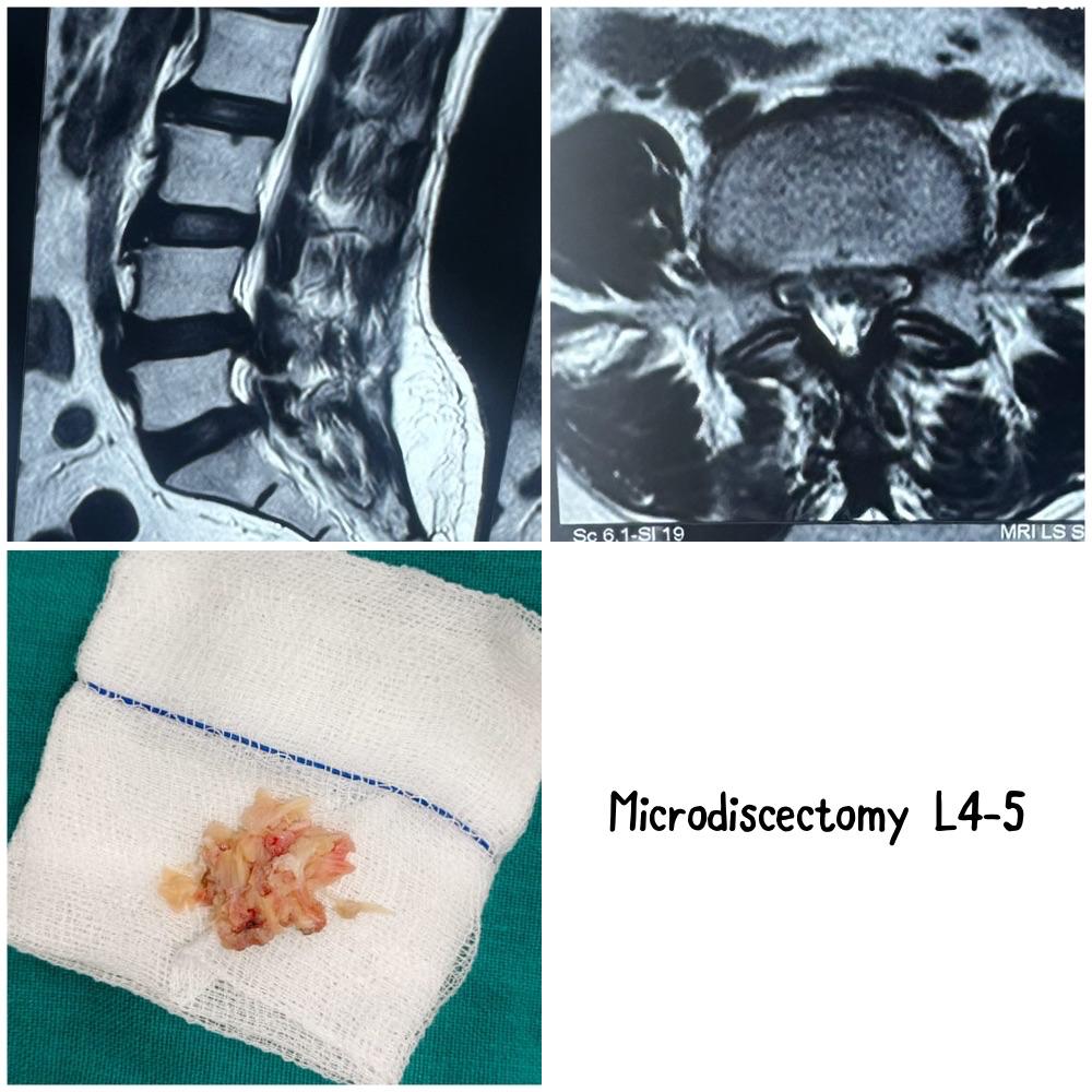
A 38-year-old software engineer presented with severe sciatic pain affecting his right leg, preventing him from working and performing daily activities. The pain had been progressively worsening over 3 months despite conservative treatment.

Initial Assessment

  • Patient: 38-year-old male, IT professional
  • Chief Complaint: Right leg sciatica, numbness, weakness
  • Duration: 3 months of progressive symptoms
  • Previous Treatment: Physical therapy, medications, rest
  • Impact: Unable to work, significant quality of life impairment

Diagnostic Evaluation

Clinical Examination

Neurological Findings:

  • Motor Weakness: Grade 4/5 in dorsiflexion of right foot
  • Sensory Changes: Decreased sensation in L5 dermatome
  • Reflexes: Diminished right patellar reflex
  • Straight Leg Raise: Positive at 30 degrees on right side

Imaging Studies

MRI Lumbar Spine revealed:

  • L4-L5 Disc Herniation: Large extruded disc fragment
  • Nerve Root Compression: Significant compression of L5 nerve root
  • Spinal Stenosis: Mild lateral recess stenosis
  • Disc Degeneration: Moderate disc desiccation

Treatment Decision

Indications for Surgery

After 3 months of failed conservative treatment, surgical intervention was recommended due to:

  • Progressive neurological deficit: Motor weakness
  • Severe pain: Uncontrolled with medications
  • Functional impairment: Inability to work
  • MRI findings: Large disc fragment causing compression

Surgical Options Discussed

  1. Microdiscectomy: Minimally invasive removal of disc fragment
  2. Endoscopic Discectomy: Alternative minimally invasive approach
  3. Open Discectomy: Traditional surgical approach

Patient elected to proceed with microdiscectomy for optimal visualization and proven outcomes.

Surgical Procedure

Pre-operative Preparation

  • Medical Clearance: Comprehensive health assessment
  • Anesthesia: General anesthesia
  • Positioning: Prone position on spinal table
  • Equipment: Microscope, surgical instruments, fluoroscopy

Surgical Technique

Approach and Exposure:

  1. Incision: 2.5 cm midline incision over L4-L5 level
  2. Dissection: Subperiosteal dissection of paraspinal muscles
  3. Laminotomy: Partial removal of lamina for access
  4. Fluoroscopy: Confirmation of correct level

Microscopic Discectomy:

  1. Microscope Setup: High-magnification visualization
  2. Epidural Space: Careful entry into epidural space
  3. Nerve Root Identification: L5 nerve root identified and protected
  4. Disc Fragment Removal: Large extruded fragment removed
  5. Disc Space: Exploration and removal of loose fragments
  6. Hemostasis: Control of bleeding

Intra-operative Findings

  • Large Fragment: 8mm extruded disc fragment
  • Nerve Compression: Significant compression of L5 root
  • Inflammation: Marked epidural inflammation
  • Disc Material: Additional loose fragments in disc space

Post-operative Care

Immediate Recovery

  • Hospital Stay: 24-hour observation
  • Pain Management: Multimodal analgesia
  • Mobilization: Ambulation on day of surgery
  • Discharge: Home with oral medications

Rehabilitation Protocol

Week 1-2:

  • Light walking as tolerated
  • No bending, twisting, or lifting
  • Incision care
  • Gradual reduction of pain medications

Week 3-6:

  • Progressive walking program
  • Core strengthening exercises
  • Gentle stretching
  • Return to light desk work

Week 6-12:

  • Advanced strengthening
  • Return to normal activities
  • Sports-specific training if applicable
  • Full activity clearance

Outcome and Results

Immediate Post-operative Results

  • Pain Relief: Immediate relief from sciatic pain
  • Motor Recovery: Improved strength in dorsiflexion
  • Sensation: Gradual return of normal sensation
  • Mobility: Ambulating without assistance

3-Month Follow-up

Clinical Assessment:

  • Pain: Complete resolution of leg pain
  • Motor: Full strength restored (5/5)
  • Sensation: Normal sensation in leg
  • Function: Returned to full work duties

Patient-Reported Outcomes:

  • Satisfaction: Very satisfied with surgical outcome
  • Quality of Life: Significant improvement
  • Return to Work: Full duties after 6 weeks
  • Activity Level: Resumed all previous activities

Key Surgical Principles

Microdiscectomy Advantages

  1. Minimally Invasive: Small incision, minimal tissue damage
  2. Direct Visualization: Magnified view of surgical field
  3. Precise Decompression: Accurate removal of pathology
  4. Rapid Recovery: Early mobilization and return to activity
  5. High Success Rate: 85-95% success rate for appropriate patients

Technical Considerations

  1. Level Confirmation: Critical to operate at correct level
  2. Nerve Protection: Prevent iatrogenic nerve injury
  3. Complete Removal: Remove all compressive fragments
  4. Hemostasis: Prevent postoperative hematoma
  5. Minimal Resection: Preserve as much normal anatomy as possible

Complications and Prevention

Potential Complications

  • Dural Tear: Cerebrospinal fluid leak
  • Nerve Injury: Temporary or permanent nerve damage
  • Infection: Surgical site infection
  • Hematoma: Postoperative bleeding
  • Recurrent Herniation: Return of disc herniation

Prevention Strategies

  • Meticulous Technique: Careful tissue handling
  • Proper Patient Selection: Appropriate candidate identification
  • Adequate Training: Surgical expertise and experience
  • Postoperative Care: Proper rehabilitation and monitoring

Long-term Results

Expected Outcomes

  • Pain Relief: 85-95% success rate
  • Return to Work: 6-12 weeks for most patients
  • Durability: Long-lasting relief in majority of cases
  • Reherniation Rate: 5-10% risk of recurrent disc herniation

Patient Selection Criteria

  • Significant Symptoms: Severe pain or neurological deficit
  • Failed Conservative: 6-12 weeks of non-surgical treatment
  • Corresponding Imaging: MRI findings match symptoms
  • Good Health: Appropriate surgical candidate

Patient Education

Pre-operative Counseling

  • Expectations: Realistic goals for recovery
  • Risks: Potential complications and outcomes
  • Rehabilitation: Importance of postoperative therapy
  • Return to Activity: Gradual return to normal activities

Post-operative Instructions

  • Activity Restrictions: Proper lifting and bending limits
  • Wound Care: Incision care and monitoring
  • Follow-up: Regular postoperative visits
  • Emergency Signs: When to seek immediate care

This case demonstrates the successful management of lumbar disc herniation using microdiscectomy, resulting in rapid pain relief and return to normal function. The minimally invasive approach provided excellent visualization while minimizing tissue trauma and recovery time.

For expert evaluation and treatment of spine conditions including disc herniations, contact VPL Ortho and Spine Clinic at +91 9042353157.

Read More
Microdiscectomy - Relief from Sciatic Pain
Awareness By Dr. Vijaya Anand S on 2026-04-18

Prevent Back Pain - Essential Tips for a Healthy Spine

Why Back Pain Matters

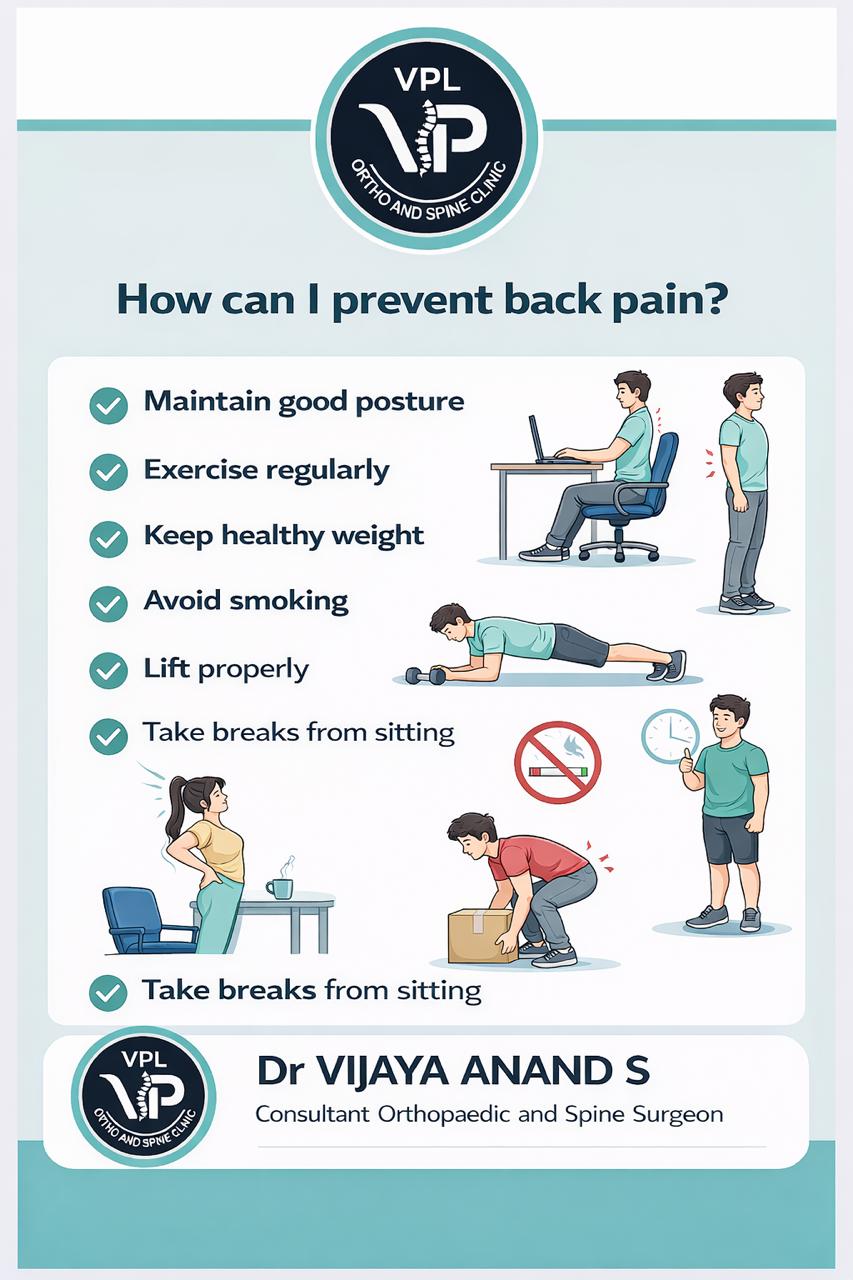
Back pain is one of the most common health problems affecting people of all ages. It can range from a dull, constant ache to a sudden, sharp pain that makes it difficult to move. The good news is that most back pain can be prevented with proper habits and lifestyle modifications.

Common Causes of Back Pain

Postural Issues:

  • Poor sitting posture at work
  • Slouching while standing or walking
  • Incorrect lifting techniques
  • Sleeping on unsupportive mattresses

Lifestyle Factors:

  • Sedentary lifestyle
  • Lack of regular exercise
  • Excess body weight
  • Smoking (reduces blood flow to spine)

Medical Conditions:

  • Herniated discs
  • Arthritis
  • Spinal stenosis
  • Muscle strains

Prevention Strategies

At Work

  • Ergonomic Setup: Adjust your chair and monitor to proper height
  • Regular Breaks: Stand and stretch every 30 minutes
  • Proper Posture: Keep your back straight and shoulders relaxed
  • Support: Use lumbar support if needed

Daily Habits

  • Exercise: Regular physical activity strengthens back muscles
  • Weight Management: Maintain healthy weight to reduce spinal stress
  • Proper Lifting: Bend at knees, not waist
  • Footwear: Wear supportive shoes

Sleep Position

  • Mattress: Use medium-firm mattress
  • Pillow: Keep neck aligned with spine
  • Position: Sleep on your side or back with proper support

Exercises for Back Health

Stretching Exercises

  1. Cat-Cow Stretch: Improves spinal flexibility
  2. Knee-to-Chest: Relieves lower back tension
  3. Child's Pose: Gentle back stretch
  4. Hamstring Stretches: Reduces back strain

Strengthening Exercises

  1. Plank: Core strengthening
  2. Bridge: Glute and lower back muscles
  3. Bird-Dog: Improves balance and core stability
  4. Partial Crunches: Abdominal strengthening

When to Seek Medical Help

Contact your orthopedic specialist if you experience:

  • Pain that persists for more than 2 weeks
  • Pain that radiates down your legs
  • Numbness or weakness in legs
  • Difficulty with bladder or bowel control
  • Pain following an injury or accident

Warning Signs Requiring Immediate Attention

  • Sudden, severe back pain
  • Pain after a fall or injury
  • Fever with back pain
  • Unexplained weight loss with back pain

Treatment Options

Depending on the cause and severity, treatment may include:

  • Physical therapy
  • Pain medications
  • Hot/cold therapy
  • In severe cases: surgical intervention

Remember, prevention is always better than cure. By adopting these healthy habits, you can significantly reduce your risk of developing back pain and maintain a healthy, active lifestyle.

For personalized assessment and treatment of back pain, visit VPL Ortho and Spine Clinic. Call +91 9042353157 for an appointment.

Read More
Prevent Back Pain - Essential Tips for a Healthy Spine
Awareness By Dr. Vijaya Anand S on 2026-04-18

Sitting Awareness - Protecting Your Spine in the Digital Age

The Sitting Epidemic

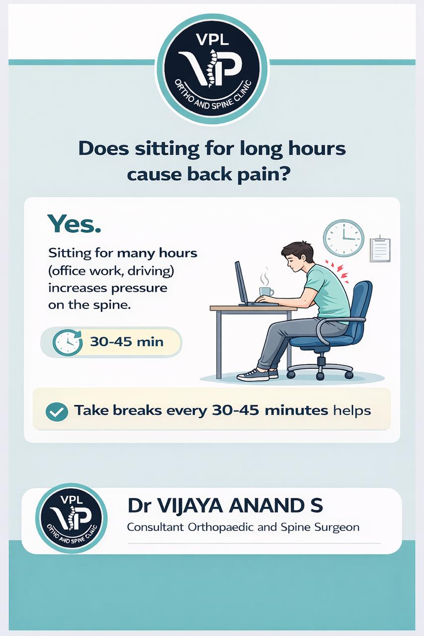
In today's digital world, most of us spend hours sitting - at desks, in meetings, commuting, or relaxing at home. This sedentary lifestyle is taking a toll on our spine health. Prolonged sitting can lead to chronic back pain, poor posture, and long-term spinal problems.

Why Sitting is Harmful

Physical Effects of Prolonged Sitting:

  • Muscle Imbalance: Weak glutes and tight hip flexors
  • Spinal Compression: Increased pressure on intervertebral discs
  • Poor Circulation: Reduced blood flow to spine and muscles
  • Postural Problems: Forward head posture and rounded shoulders
  • Metabolic Issues: Increased risk of diabetes and heart disease

Ergonomic Sitting Guidelines

Proper Desk Setup

  • Monitor Height: Top of screen at or below eye level
  • Chair Height: Feet flat on floor, knees at 90-degree angle
  • Keyboard Position: Elbows at 90 degrees, wrists straight
  • Mouse Distance: Close to keyboard, avoid reaching
  • Document Holder: At same height as monitor

Chair Adjustments

  • Lumbar Support: Adjust to support natural curve of lower back
  • Seat Depth: 2-3 finger gap between knees and seat edge
  • Armrests: Shoulders relaxed, elbows at 90 degrees
  • Backrest Angle: Slightly reclined (100-110 degrees)

Movement Strategies

The 30-30 Rule

  • Every 30 minutes: Stand up and move for 30 seconds
  • Every 2 hours: Take a 5-minute walking break
  • Micro-breaks: Stand, stretch, or change position frequently

Desk Exercises

  1. Neck Stretches: Gently tilt head side to side
  2. Shoulder Rolls: Forward and backward shoulder circles
  3. Spinal Twists: Gentle torso rotations while seated
  4. Hip Flexor Stretch: Stand and gently stretch hips
  5. Wrist Stretches: Extend and flex wrists regularly

Standing Desks

  • Adjustable Desks: Alternate between sitting and standing
  • Anti-Fatigue Mats: Reduce strain when standing
  • Proper Height: Elbows at 90 degrees when standing
  • Foot Support: Use footrest if needed

Posture Correction

Ideal Sitting Posture

  • Head: Aligned with shoulders, not forward
  • Shoulders: Relaxed, not hunched
  • Back: Straight with natural lumbar curve
  • Hips: Slightly higher than knees
  • Feet: Flat on floor or footrest

Common Posture Mistakes

  • Forward Head: "Text neck" from phone/computer use
  • Slumped Shoulders: Rounded upper back
  • Crossed Legs: Can cause pelvic imbalance
  • Leaning Forward: Strains back and neck muscles

Lifestyle Modifications

Outside of Work

  • Regular Exercise: Counteract sitting with physical activity
  • Stretching: Daily flexibility routine
  • Core Strengthening: Support your spine
  • Active Hobbies: Choose movement-based activities

Daily Habits

  • Take the stairs: Instead of elevators
  • Walk during calls: Pace while on phone
  • Park further away: Extra steps to your destination
  • Active breaks: Walk during lunch breaks

Warning Signs

Seek medical attention if you experience:

  • Persistent back pain lasting more than a week
  • Pain that radiates to legs or arms
  • Numbness or tingling in extremities
  • Weakness in legs or arms
  • Difficulty with daily activities

Technology Solutions

Ergonomic Tools

  • Monitor Arms: Adjustable screen positioning
  • Ergonomic Keyboards: Reduce wrist strain
  • Vertical Mice: Natural hand position
  • Lumbar Cushions: Additional back support
  • Footrests: Proper leg positioning

Apps and Reminders

  • Break Reminders: Apps that prompt movement
  • Posture Trackers: Devices that monitor sitting habits
  • Ergonomic Assessments: Online tools for workspace setup

Long-Term Health Impact

Benefits of Proper Sitting Habits:

  • Reduced back and neck pain
  • Improved posture and appearance
  • Increased energy and productivity
  • Better overall health
  • Prevention of chronic conditions

Remember, your body is designed to move. While modern life requires sitting, being mindful about how you sit and incorporating regular movement can protect your spine and overall health.

For personalized ergonomic advice and treatment of sitting-related pain, visit VPL Ortho and Spine Clinic. Call +91 9042353157 to schedule an appointment.

Read More
Sitting Awareness - Protecting Your Spine in the Digital Age
Surgery Case By Dr. Vijaya Anand S on 2026-04-18

Total Knee Replacement - Restoring Mobility and Quality of Life

Case Presentation

A 68-year-old retired bank manager presented with severe knee pain that had progressively worsened over 8 years, significantly limiting her ability to walk, climb stairs, and enjoy her retirement activities.

Initial Assessment

  • Patient: 68-year-old female, retired bank manager
  • Chief Complaint: Right knee pain, swelling, stiffness
  • Duration: 8 years of progressive symptoms
  • Functional Impact: Unable to walk more than 50 meters, difficulty with stairs
  • Previous Treatment: NSAIDs, physical therapy, viscosupplementation injections

Diagnostic Evaluation

Clinical Examination

Physical Findings:

  • Gait: Antalgic gait with visible limp
  • Range of Motion: Limited flexion (90 degrees) and extension (5 degrees flexion contracture)
  • Joint Stability: Medial and lateral laxity present
  • Deformity: Varus deformity of 15 degrees
  • Effusion: Moderate joint effusion present
  • Tenderness: Medial joint line tenderness

Imaging Studies

X-ray Right Knee:

  • Joint Space: Complete loss of medial joint space
  • Osteophytes: Large osteophytes medially and laterally
  • Subchondral Sclerosis: Marked sclerosis of medial tibial plateau
  • Deformity: Varus malalignment of 15 degrees
  • Bone Quality: Good bone stock, no significant osteoporosis

MRI Knee (if indicated):

  • Cartilage: Complete loss of articular cartilage medially
  • Meniscus: Complex tear of medial meniscus
  • Ligaments: Intact ACL and PCL
  • Bone Marrow Edema: Extensive bone marrow changes

Treatment Decision

Indications for Surgery

Total knee replacement was recommended based on:

  • Severe Pain: Uncontrolled with conservative measures
  • Functional Limitation: Significant impact on daily activities
  • Radiological Evidence: End-stage osteoarthritis
  • Failed Conservative: Adequate trial of non-surgical treatment
  • Quality of Life: Significant reduction in quality of life

Pre-operative Planning

Implant Selection:

  • Type: Posterior-stabilized total knee prosthesis
  • Size: Based on pre-operative measurements
  • Material: Cobalt-chromium femoral component, polyethylene insert
  • Fixation: Cemented fixation for immediate stability

Surgical Procedure

Pre-operative Preparation

  • Medical Clearance: Comprehensive cardiac and medical evaluation
  • Anesthesia: Spinal anesthesia with sedation
  • Positioning: Supine position with tourniquet application
  • Equipment: Total knee arthroplasty instrument set, computer navigation

Surgical Technique

Approach and Exposure:

  1. Incision: Midline anterior incision (15 cm)
  2. Arthrotomy: Medial parapatellar arthrotomy
  3. Patellar Management: Patellar eversion and preparation
  4. Exposure: Complete exposure of distal femur and proximal tibia

Bone Preparation:

  1. Distal Femur: Intramedullary guide for distal femoral cut
  2. Proximal Tibia: Extramedullary guide for tibial resection
  3. Patella: Resection of patellar surface
  4. Sizing: Appropriate sizing of femoral and tibial components

Component Implantation:

  1. Trial Components: Trial implants for assessment
  2. Range of Motion: Assessment of flexion, extension, stability
  3. Final Components: Cementation of femoral, tibial, and patellar components
  4. Polyethylene Insert: Appropriate thickness insert placement

Intra-operative Findings

  • Cartilage: Complete loss of articular cartilage
  • Bone Quality: Good bone stock for cement fixation
  • Deformity: Significant varus deformity corrected
  • Stability: Achieved excellent stability with prosthesis

Post-operative Care

Immediate Recovery

  • Hospital Stay: 4 days for monitoring and rehabilitation
  • Pain Management: Multimodal analgesia protocol
  • Blood Loss: Minimal blood loss, no transfusion required
  • Mobilization: Ambulation on postoperative day 1 with walker

Rehabilitation Protocol

Week 1-2:

  • Weight Bearing: Full weight bearing as tolerated
  • Range of Motion: Goal of 90 degrees flexion
  • Strengthening: Quadriceps and hamstring exercises
  • Gait Training: Walker to cane progression

Week 3-6:

  • Range of Motion: Goal of 110-120 degrees flexion
  • Strengthening: Advanced strengthening program
  • Functional Activities: Stair climbing, sit-to-stand exercises
  • Walking: Progress to independent ambulation

Week 6-12:

  • Range of Motion: Full functional range of motion
  • Activities: Return to light recreational activities
  • Driving: Clearance for driving (typically 6-8 weeks)
  • Sports: Low-impact sports as tolerated

Outcome and Results

3-Month Follow-up

Clinical Assessment:

  • Pain: Complete resolution of arthritic pain
  • Range of Motion: 115 degrees flexion, full extension
  • Stability: Stable knee with no laxity
  • Function: Walking unlimited distance, stair climbing without difficulty

Radiological Assessment:

  • Component Position: Optimal positioning of all components
  • Alignment: Neutral mechanical alignment achieved
  • Fixation: No signs of loosening or subsidence
  • Bone-Cement Interface: Complete incorporation

6-Month Follow-up

Patient-Reported Outcomes:

  • Pain Score: Reduced from 8/10 to 1/10
  • Functional Score: Significant improvement in knee scores
  • Quality of Life: Return to most desired activities
  • Satisfaction: Very satisfied with surgical outcome

Functional Achievements:

  • Walking: Unlimited walking distance
  • Stairs: Able to climb stairs normally
  • Activities: Return to gardening, walking, light sports
  • Independence: Full independence in daily activities

Key Surgical Principles

Total Knee Replacement Success Factors

  1. Proper Alignment: Mechanical axis restoration
  2. Component Positioning: Optimal femoral and tibial positioning
  3. Soft Tissue Balance: Equal flexion and extension gaps
  4. Patellar Tracking: Proper patellar alignment
  5. Fixation: Secure cement fixation

Technical Considerations

  1. Bone Cuts: Precise bone resection angles
  2. Soft Tissue: Adequate release of tight structures
  3. Rotation: Proper rotational alignment of components
  4. Sizing: Appropriate component sizing
  5. Range of Motion: Achieve functional range of motion

Complications and Prevention

Potential Complications

  • Infection: Prosthetic joint infection
  • DVT/PE: Deep vein thrombosis or pulmonary embolism
  • Stiffness: Arthrofibrosis limiting motion
  • Loosening: Aseptic loosening of components
  • Neurovascular Injury: Nerve or blood vessel injury

Prevention Strategies

  • Antibiotics: Prophylactic antibiotic administration
  • DVT Prophylaxis: Mechanical and chemical prophylaxis
  • Early Mobilization: Prevent stiffness and complications
  • Proper Technique: Meticulous surgical execution
  • Patient Selection: Appropriate candidate identification

Long-term Results

Expected Outcomes

  • Survivorship: 95% survivorship at 15 years
  • Pain Relief: 90% success rate for pain reduction
  • Function: Significant improvement in knee function
  • Durability: Long-lasting results with proper technique

Lifestyle Modifications

  • Activity Recommendations: Low-impact activities preferred
  • Weight Management: Maintain healthy body weight
  • Regular Exercise: Strengthening and flexibility exercises
  • Follow-up: Regular orthopedic follow-up visits

Patient Education

Pre-operative Counseling

  • Expectations: Realistic goals for recovery
  • Risks: Potential complications and their management
  • Rehabilitation: Importance of postoperative therapy
  • Long-term Care: Lifetime care of prosthetic joint

Post-operative Instructions

  • Activity Progression: Gradual return to activities
  • Wound Care: Proper incision care and monitoring
  • Warning Signs: When to seek immediate medical attention
  • Long-term Follow-up: Regular monitoring schedule

Success Metrics

Clinical Success Indicators

  • Pain Relief: Significant reduction in knee pain
  • Functional Improvement: Return to desired activities
  • Range of Motion: Functional range of motion achieved
  • Patient Satisfaction: High patient satisfaction scores

Radiological Success Indicators

  • Component Position: Optimal positioning maintained
  • Alignment: Neutral alignment preserved
  • Fixation: No evidence of loosening
  • Bone Quality: Good bone maintenance around components

This case demonstrates successful management of end-stage knee osteoarthritis through total knee replacement surgery. The outcome highlights the importance of proper patient selection, meticulous surgical technique, and comprehensive rehabilitation for optimal long-term results.

For expert evaluation and treatment of knee arthritis and joint replacement, contact VPL Ortho and Spine Clinic at +91 9042353157.

Read More
Total Knee Replacement - Restoring Mobility and Quality of Life
Awareness By Dr. Vijaya Anand S on 2026-04-18

Understanding Osteoporosis - Prevention and Management

What is Osteoporosis?

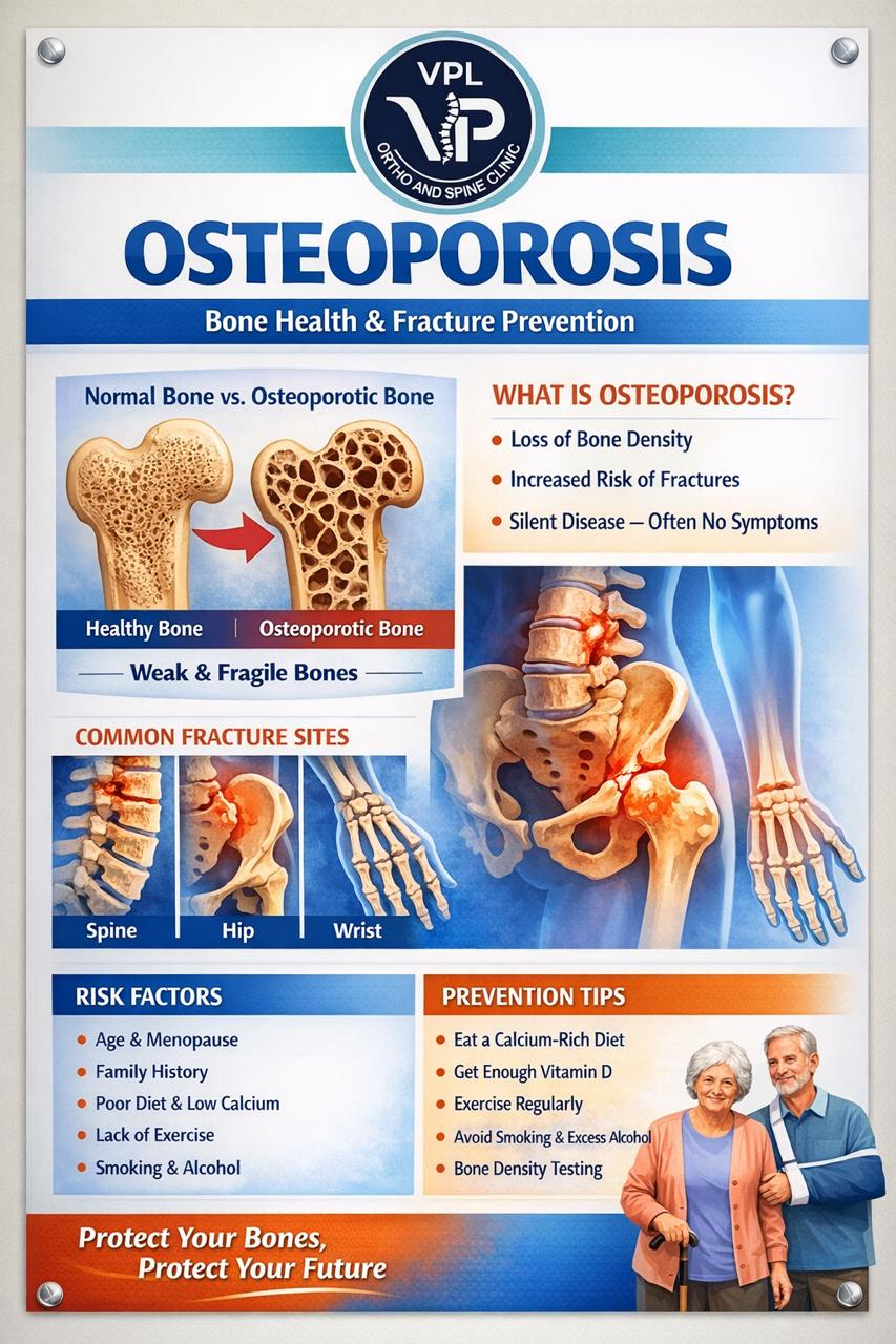
Osteoporosis is a silent bone disease that weakens bones, making them fragile and more likely to break. It occurs when the body loses too much bone, makes too little bone, or both. This condition affects millions of people worldwide, particularly women after menopause and older adults.

Risk Factors

Major risk factors include:

  • Age (risk increases after 50)
  • Female gender (especially post-menopausal)
  • Family history of osteoporosis
  • Small, thin body frame
  • Low calcium and vitamin D intake
  • Sedentary lifestyle
  • Smoking and excessive alcohol consumption
  • Certain medications (steroids, thyroid medications)

Prevention Strategies

Nutrition

  • Calcium: Adults need 1000-1200mg daily

    • Dairy products (milk, cheese, yogurt)
    • Leafy green vegetables (spinach, kale)
    • Fortified foods
    • Calcium supplements if needed
  • Vitamin D: Essential for calcium absorption

    • Sunlight exposure (15-20 minutes daily)
    • Fatty fish (salmon, mackerel)
    • Fortified milk and cereals
    • Supplements (800-1000 IU daily)

Exercise

  • Weight-bearing exercises: Walking, jogging, dancing
  • Strength training: 2-3 times per week
  • Balance exercises: Tai chi, yoga to prevent falls

Lifestyle Modifications

  • Quit smoking
  • Limit alcohol to 1-2 drinks per day
  • Maintain healthy body weight
  • Fall prevention measures at home

When to See a Doctor

Consult your orthopedic specialist if you:

  • Have a family history of osteoporosis
  • Experience height loss
  • Develop back pain without obvious cause
  • Have fractures from minor injuries
  • Are over 50 and haven't been screened

Treatment Options

If diagnosed with osteoporosis, treatment may include:

  • Prescription medications (bisphosphonates, denosumab)
  • Calcium and vitamin D supplements
  • Physical therapy
  • Lifestyle modifications

Remember, osteoporosis is preventable and manageable. Early detection and intervention can significantly reduce your risk of fractures and maintain bone health.

For personalized assessment and treatment, contact VPL Ortho and Spine Clinic at +91 9042353157.

Read More
Understanding Osteoporosis - Prevention and Management
Awareness By Dr. Vijaya Anand S on 2026-04-18

Vitamin D - The Sunshine Vitamin for Bone Health

Understanding Vitamin D

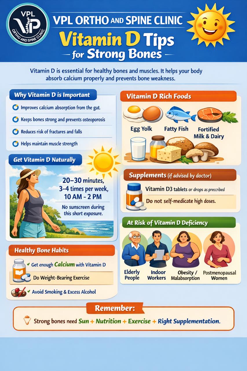
Vitamin D, often called the "sunshine vitamin," is a fat-soluble vitamin that plays a crucial role in bone health and overall wellness. Unlike other vitamins, our body can produce vitamin D when exposed to sunlight, making it unique among essential nutrients.

Why Vitamin D Matters for Bone Health

Key Functions:

  • Calcium Absorption: Helps your body absorb calcium from food
  • Bone Mineralization: Essential for building strong bones
  • Muscle Function: Supports muscle strength and balance
  • Immune Support: Boosts immune system function
  • Inflammation Reduction: Helps control inflammatory responses

Vitamin D Deficiency: A Silent Epidemic

Common Risk Factors:

  • Limited sun exposure (indoor lifestyle, office work)
  • Darker skin tones (less efficient vitamin D production)
  • Older age (skin becomes less efficient at producing vitamin D)
  • Certain medical conditions (celiac disease, Crohn's disease)
  • Obesity (vitamin D gets sequestered in fat tissue)
  • Certain medications (anti-seizure drugs, steroids)

Symptoms of Deficiency:

  • Bone pain and muscle weakness
  • Fatigue and tiredness
  • Depression or mood changes
  • Slow wound healing
  • Hair loss
  • Increased susceptibility to infections

Sources of Vitamin D

Sunlight Exposure

  • Best Source: 15-20 minutes of midday sun exposure
  • Optimal Time: 10 AM to 3 PM
  • Skin Exposure: Face, arms, legs without sunscreen
  • Seasonal Variation: Winter months reduce vitamin D production
  • Geographic Factors: Less UVB in northern latitudes

Food Sources

Natural Sources:

  • Fatty fish (salmon, mackerel, sardines)
  • Egg yolks
  • Beef liver
  • Cheese

Fortified Foods:

  • Milk and dairy products
  • Orange juice
  • Breakfast cereals
  • Plant-based milk alternatives

Supplements

  • Vitamin D3: More effective than D2
  • Dosage: 800-2000 IU daily for most adults
  • Prescription: Higher doses available for deficiency
  • Absorption: Take with fat-containing meal

Recommended Daily Intake

Age Groups:

  • Infants 0-12 months: 400 IU
  • Children 1-18 years: 600 IU
  • Adults 19-70 years: 600 IU
  • Adults 71+ years: 800 IU
  • Pregnant/Breastfeeding: 600 IU

Higher Doses May Be Needed For:

  • Obesity
  • Malabsorption conditions
  • Certain medications
  • Dark skin pigmentation
  • Limited sun exposure

Testing and Diagnosis

When to Test:

  • Risk factors for deficiency
  • Symptoms of low vitamin D
  • Bone density concerns
  • Before starting high-dose supplements

Blood Test:

  • 25-hydroxyvitamin D (25(OH)D)
  • Levels below 20 ng/mL indicate deficiency
  • Levels 21-29 ng/mL indicate insufficiency
  • Levels 30+ ng/mL are considered sufficient

Vitamin D and Bone Conditions

Osteoporosis

  • Prevention: Adequate vitamin D reduces fracture risk
  • Treatment: Essential component of osteoporosis management
  • Synergy: Works with calcium for optimal bone health

Osteomalacia

  • Adult Condition: Softening of bones due to vitamin D deficiency
  • Symptoms: Bone pain, muscle weakness, increased fracture risk
  • Treatment: High-dose vitamin D supplementation

Rickets

  • Childhood Condition: Improper bone mineralization
  • Prevention: Adequate vitamin D during pregnancy and childhood
  • Treatment: Vitamin D and calcium supplementation

Special Considerations

Pregnancy and Breastfeeding

  • Fetal Development: Critical for baby's bone formation
  • Maternal Health: Prevents pregnancy complications
  • Breast Milk: Vitamin D content depends on maternal levels

Elderly Population

  • Fall Prevention: Reduces fall risk through muscle strength
  • Fracture Protection: Decreases hip fracture risk
  • Cognitive Health: May support brain function

Chronic Conditions

  • Diabetes: May improve insulin sensitivity
  • Heart Disease: Potential cardiovascular benefits
  • Autoimmune Disorders: May modulate immune responses

Safety and Toxicity

Upper Safe Limits:

  • Adults: 4,000 IU daily
  • Children: Varies by age
  • Toxicity: Rare, usually from excessive supplementation

Symptoms of Excess:

  • Nausea and vomiting
  • Constipation
  • Weakness
  • Kidney stones
  • High blood calcium levels

Practical Tips for Optimal Vitamin D

Daily Habits

  • Morning Sun: 10-15 minutes of morning sun exposure
  • Outdoor Activities: Regular outdoor exercise
  • Window Time: Sit near windows for indirect sun exposure
  • Diet: Include vitamin D-rich foods regularly

Seasonal Strategies

  • Summer: Build up vitamin D stores
  • Winter: Consider supplementation
  • Cloudy Days: May need extra supplementation
  • High Latitudes: Year-round supplementation often needed

When to Consult Your Doctor

Seek medical advice if you:

  • Have symptoms of vitamin D deficiency
  • Have risk factors for deficiency
  • Are considering high-dose supplements
  • Have chronic medical conditions
  • Are pregnant or breastfeeding
  • Have been diagnosed with osteoporosis

Remember, vitamin D is essential for bone health and overall wellness. Maintaining adequate levels through sun exposure, diet, and supplementation when necessary can help prevent bone problems and support your overall health.

For personalized vitamin D assessment and bone health evaluation, visit VPL Ortho and Spine Clinic. Call +91 9042353157 to schedule your appointment.

Read More
Vitamin D - The Sunshine Vitamin for Bone Health
Awareness By Dr. Vijaya Anand on 2026-04-16

Welcome to VPL Ortho and Spine Clinic Blog

Welcome to our blog! Here at VPL Ortho and Spine Clinic, we are committed to educating our patients and the community about orthopedic health, spine care, and preventive measures.

What You'll Find Here

  • Surgery Case Studies: Detailed accounts of successful surgeries we've performed, showcasing our expertise and the positive outcomes our patients achieve
  • Awareness Posts: Information about common orthopedic conditions, preventive care tips, and public health awareness
  • Treatment Updates: Latest advancements in orthopedic and spine treatments
  • Recovery Tips: Guidance on post-surgery care and rehabilitation

Our goal is to empower you with knowledge so you can make informed decisions about your orthopedic health. Whether you're dealing with chronic pain, recovering from an injury, or simply want to maintain healthy joints and spine, our blog is here to help.

Stay tuned for regular updates from our experienced orthopedic specialists!

For appointments or consultations, contact us at +91 9042353157.

Read More