Case Presentation
A 38-year-old software engineer presented with severe sciatic pain affecting his right leg, preventing him from working and performing daily activities. The pain had been progressively worsening over 3 months despite conservative treatment.
Initial Assessment
- Patient: 38-year-old male, IT professional
- Chief Complaint: Right leg sciatica, numbness, weakness
- Duration: 3 months of progressive symptoms
- Previous Treatment: Physical therapy, medications, rest
- Impact: Unable to work, significant quality of life impairment
Diagnostic Evaluation
Clinical Examination
Neurological Findings:
- Motor Weakness: Grade 4/5 in dorsiflexion of right foot
- Sensory Changes: Decreased sensation in L5 dermatome
- Reflexes: Diminished right patellar reflex
- Straight Leg Raise: Positive at 30 degrees on right side
Imaging Studies
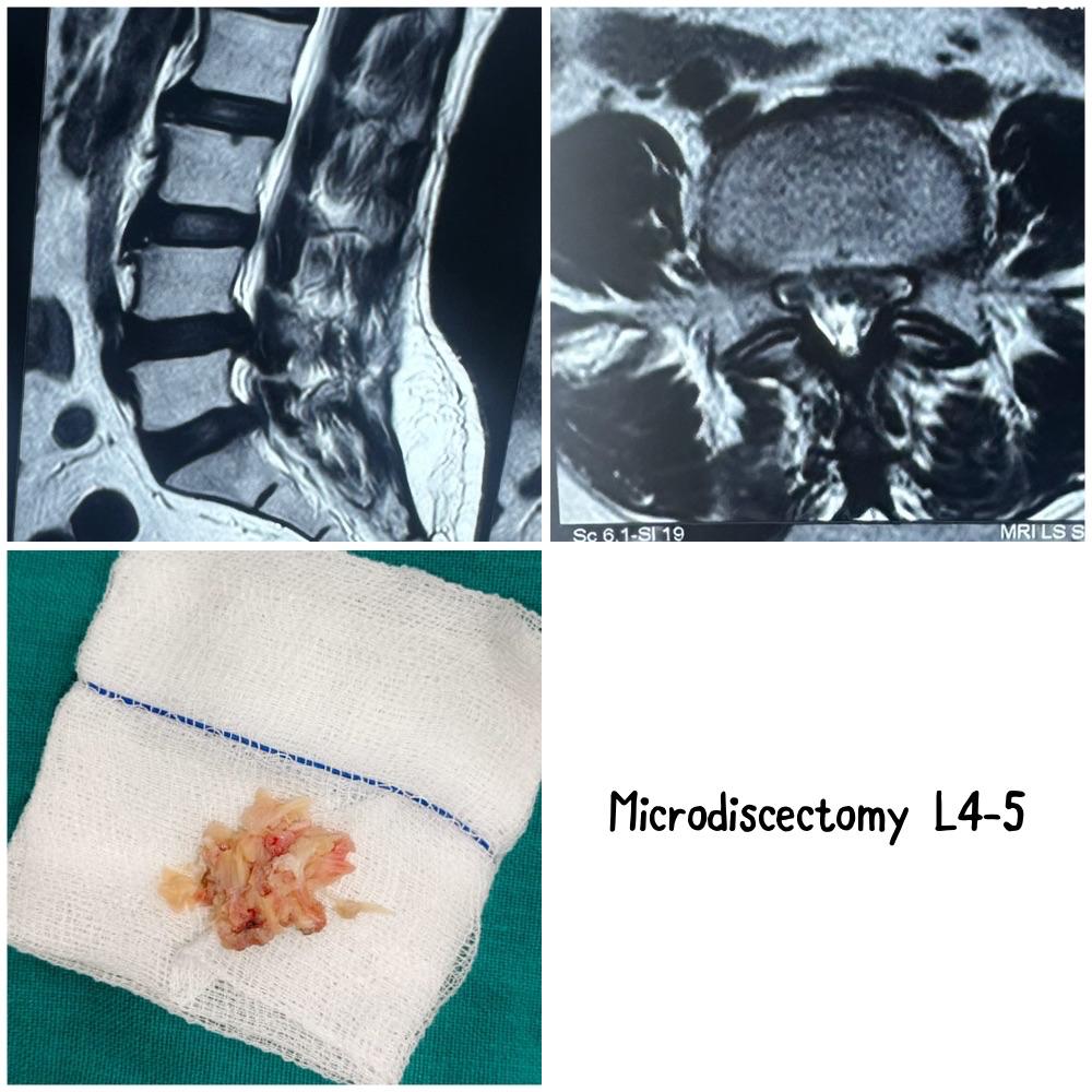
MRI Lumbar Spine revealed:
- L4-L5 Disc Herniation: Large extruded disc fragment
- Nerve Root Compression: Significant compression of L5 nerve root
- Spinal Stenosis: Mild lateral recess stenosis
- Disc Degeneration: Moderate disc desiccation
Treatment Decision
Indications for Surgery
After 3 months of failed conservative treatment, surgical intervention was recommended due to:
- Progressive neurological deficit: Motor weakness
- Severe pain: Uncontrolled with medications
- Functional impairment: Inability to work
- MRI findings: Large disc fragment causing compression
Surgical Options Discussed
- Microdiscectomy: Minimally invasive removal of disc fragment
- Endoscopic Discectomy: Alternative minimally invasive approach
- Open Discectomy: Traditional surgical approach
Patient elected to proceed with microdiscectomy for optimal visualization and proven outcomes.
Surgical Procedure
Pre-operative Preparation
- Medical Clearance: Comprehensive health assessment
- Anesthesia: General anesthesia
- Positioning: Prone position on spinal table
- Equipment: Microscope, surgical instruments, fluoroscopy
Surgical Technique
Approach and Exposure:
- Incision: 2.5 cm midline incision over L4-L5 level
- Dissection: Subperiosteal dissection of paraspinal muscles
- Laminotomy: Partial removal of lamina for access
- Fluoroscopy: Confirmation of correct level
Microscopic Discectomy:
- Microscope Setup: High-magnification visualization
- Epidural Space: Careful entry into epidural space
- Nerve Root Identification: L5 nerve root identified and protected
- Disc Fragment Removal: Large extruded fragment removed
- Disc Space: Exploration and removal of loose fragments
- Hemostasis: Control of bleeding
Intra-operative Findings
- Large Fragment: 8mm extruded disc fragment
- Nerve Compression: Significant compression of L5 root
- Inflammation: Marked epidural inflammation
- Disc Material: Additional loose fragments in disc space
Post-operative Care
Immediate Recovery
- Hospital Stay: 24-hour observation
- Pain Management: Multimodal analgesia
- Mobilization: Ambulation on day of surgery
- Discharge: Home with oral medications
Rehabilitation Protocol
Week 1-2:
- Light walking as tolerated
- No bending, twisting, or lifting
- Incision care
- Gradual reduction of pain medications
Week 3-6:
- Progressive walking program
- Core strengthening exercises
- Gentle stretching
- Return to light desk work
Week 6-12:
- Advanced strengthening
- Return to normal activities
- Sports-specific training if applicable
- Full activity clearance
Outcome and Results
Immediate Post-operative Results
- Pain Relief: Immediate relief from sciatic pain
- Motor Recovery: Improved strength in dorsiflexion
- Sensation: Gradual return of normal sensation
- Mobility: Ambulating without assistance
3-Month Follow-up
Clinical Assessment:
- Pain: Complete resolution of leg pain
- Motor: Full strength restored (5/5)
- Sensation: Normal sensation in leg
- Function: Returned to full work duties
Patient-Reported Outcomes:
- Satisfaction: Very satisfied with surgical outcome
- Quality of Life: Significant improvement
- Return to Work: Full duties after 6 weeks
- Activity Level: Resumed all previous activities
Key Surgical Principles
Microdiscectomy Advantages
- Minimally Invasive: Small incision, minimal tissue damage
- Direct Visualization: Magnified view of surgical field
- Precise Decompression: Accurate removal of pathology
- Rapid Recovery: Early mobilization and return to activity
- High Success Rate: 85-95% success rate for appropriate patients
Technical Considerations
- Level Confirmation: Critical to operate at correct level
- Nerve Protection: Prevent iatrogenic nerve injury
- Complete Removal: Remove all compressive fragments
- Hemostasis: Prevent postoperative hematoma
- Minimal Resection: Preserve as much normal anatomy as possible
Complications and Prevention
Potential Complications
- Dural Tear: Cerebrospinal fluid leak
- Nerve Injury: Temporary or permanent nerve damage
- Infection: Surgical site infection
- Hematoma: Postoperative bleeding
- Recurrent Herniation: Return of disc herniation
Prevention Strategies
- Meticulous Technique: Careful tissue handling
- Proper Patient Selection: Appropriate candidate identification
- Adequate Training: Surgical expertise and experience
- Postoperative Care: Proper rehabilitation and monitoring
Long-term Results
Expected Outcomes
- Pain Relief: 85-95% success rate
- Return to Work: 6-12 weeks for most patients
- Durability: Long-lasting relief in majority of cases
- Reherniation Rate: 5-10% risk of recurrent disc herniation
Patient Selection Criteria
- Significant Symptoms: Severe pain or neurological deficit
- Failed Conservative: 6-12 weeks of non-surgical treatment
- Corresponding Imaging: MRI findings match symptoms
- Good Health: Appropriate surgical candidate
Patient Education
Pre-operative Counseling
- Expectations: Realistic goals for recovery
- Risks: Potential complications and outcomes
- Rehabilitation: Importance of postoperative therapy
- Return to Activity: Gradual return to normal activities
Post-operative Instructions
- Activity Restrictions: Proper lifting and bending limits
- Wound Care: Incision care and monitoring
- Follow-up: Regular postoperative visits
- Emergency Signs: When to seek immediate care
This case demonstrates the successful management of lumbar disc herniation using microdiscectomy, resulting in rapid pain relief and return to normal function. The minimally invasive approach provided excellent visualization while minimizing tissue trauma and recovery time.
For expert evaluation and treatment of spine conditions including disc herniations, contact VPL Ortho and Spine Clinic at +91 9042353157.